Overview of Osteoarthritis and Physiotherapy Management
Osteoarthritis is the most common form of arthritis, affecting millions of people worldwide. It occurs when the protective cartilage that cushions the ends of your bones wears down over time. Although osteoarthritis can damage any joint, the disorder most commonly affects joints in your hands, knees, hips and spine.
Osteoarthritis symptoms can usually be managed, although the damage to joints can't be reversed. Staying active, maintaining a healthy weight and some treatments might slow progression of the disease and help improve pain and joint function.
Osteoarthritis symptoms often develop slowly and worsen over time. Signs and symptoms of osteoarthritis include:
- Pain. Affected joints might hurt during or after movement.
- Stiffness. Joint stiffness might be most noticeable upon awakening or after being inactive.
- Tenderness. Your joint might feel tender when you apply light pressure to or near it.
- Loss of flexibility. You might not be able to move your joint through its full range of motion.
- Grating sensation. You might feel a grating sensation when you use the joint, and you might hear popping or crackling.
- Bone spurs. These extra bits of bone, which feel like hard lumps, can form around the affected joint.
- Swelling. This might be caused by soft tissue inflammation around the joint.
Knee osteoarthritis (OA), also known as degenerative joint disease, is typically the result of wear and tear and progressive loss of articular cartilage. It is most common in elderly people and can be divided into two types, primary and secondary:
- Primary osteoarthritis - is articular degeneration without any apparent underlying cause.
- Secondary osteoarthritis - is the consequence of either an abnormal concentration of force across the joint as with post-traumatic causes or abnormal articular cartilage, such as rheumatoid arthritis (RA).
Although osteoarthritis can get worse over time, it is not an inevitable part of ageing and isn’t always progressive (Australian Commission on Safety and Quality in Health Care, 2017). You can manage your symptoms and improve your physical activity by revising risk factors such as excessive weight. Being overweight increases risk of osteoarthritis by two times, being obese increases the risk by four times (Australian Commission on Safety and Quality in Health Care, 2017).
Osteoarthritis isn’t necessarily degenerative, and it doesn’t always get worse over time despite common belief. Osteoarthritis can be made worse by avoiding movement and exercise. For a long time, the recommendation has been to “rest, avoid pain” however the associated muscle wastage and decrease in strength and mobility of the affected joint is what makes osteoarthritis appear to be degenerative.
Osteoarthritis is typically a progressive disease that may eventually lead to disability. The intensity of the clinical symptoms may vary from each individual. However, they typically become more severe, more frequent, and more debilitating over time. The rate of progression also varies for each individual.
The Center for Disease Control and Prevention has found that the number of people suffering from knee pain disorder is gradually rising, with approximately 1 in 2 people likely to develop symptomatic knee OA in their lifetime leading to significant impact on health, workplace productivity and economic costs.
OA pain is easily identifiable through diagnostics and common symptoms. Some people who suffer from immense osteoarthritis knee pain may only show mild changes on x-ray, so it is extremely important to concentrate on the symptoms, rather than just the x-rays. Here is a look at the stages of osteoarthritis of the knee ranging from normal, minor, mild, moderate and severe stages, with appropriate treatment plans.
Stage 0 – Normal
When the knee shows no signs of osteoarthritis, it is classified as Stage 0, which is normal knee health, with no known impairment or signs of joint damage.
Stage 1 – Minor
Stage 1 OA patients will develop very minor wear & tear and bone spur growths at the end of the knee joints. However, at this stage it is unlikely you will feel pain or discomfort.
Stage 2 – Mild
In Stage 2, diagnostic images or X-rays of knee joints will show more bone spur growth, and though the space between the bones appear normal, people will begin experiencing symptoms of joint pain. Typically, the area around the knee joints will feel stiff and uncomfortable, particularly when sitting for an extended period, after rising in the morning, or after a workout. Though the cartilage and soft tissues remains at a healthy size, there is proteolytic breakdown of the cartilage matrix from an increased production of enzymes, such as metalloproteinases.
Stage 3 – Moderate
Stage 3 is referred to as “moderate”, where there is obvious erosion to the cartilage surface between bones and fibrillation narrows the gap between the bones. There are proteoglycan and collagen fragments released into the synovial fluid as the disease progresses, wherein the bones develop spurs at the joints as it becomes rougher. With the progression of osteoarthritis of the knee, there is obvious joint inflammation which causes frequent pain when walking, running, squatting, extending or kneeling. Along with joint stiffness after sitting for long or when waking up in the morning, there may be popping or snapping sounds when walking.
Stage 4 – Severe
Stage 4 is considered to be severe. In stage 4 the joint space between the bones are considerably reduced, causing the cartilage to wear off, leaving the joint stiff. The breakdown of cartilage leads to a chronic inflammatory response, with decreased synovial fluid that causes friction, greater pain and discomfort when walking or moving the joint.
There is increased production of synovial metalloproteinases, cytokines and TNF that can diffuse back into the cartilage to destroy soft tissue around the knee. The advanced stage of the disease shows development of more spurs causing excruciating pain, which makes even everyday chores, including walking and descending stairs a challenge.
In cases of severe OA of the knee, an option is performing osteotomy or bone realignment surgery, wherein the orthopedic surgeon cuts the bone above or below the knee to shorten the length and help realign it for less stress on the knee joint. This surgery helps protect the knee by shifting the weight of the body away from the site of the bone spur growth and bone damage.
Another surgical option is total knee replacement, or arthroplasty. During this surgical procedure, the damaged joint is removed and replaced with a plastic or metal prosthesis device. Recovery from surgery may take several weeks and requires patience and discipline, with continuous physical and occupational therapy to regain full mobility.
Treatment/Management
Treatment for knee OA can be broken down into conservative and surgical management.
Initial treatment always begins with conservative modalities and moves to surgical treatment once conservative management has been exhausted. There is a wide range of conservative modalities is available for the treatment of knee OA. These interventions do not alter the underlying disease process, but their goal is to reduce pain and optimise function for as long as possible.
Conservative Treatment Options
The primary treatment for OA knee conservatively is exercise therapy within physiotherapy. Physiotherapy normally involves
- Patient education
- Exercise therapy
- Activity modification
- Advice on weight loss
- Knee bracing
The first-line treatment for all patients with symptomatic knee osteoarthritis includes patient education and physiotherapy. A combination of supervised exercises and a home exercise program have been shown to have the best results. These benefits are lost after 6 months if the exercises are stopped.
Weight loss is valuable in all stages of knee OA. It is indicated in patients with symptomatic OA with a body mass index greater than 25. The best recommendation to achieve weight loss is with diet control and low-impact aerobic exercise.
Knee bracing in OA can be used. Offloading-type braces which shift the load away from the involved knee compartment. This can be effective when there is a valgus or varus deformity.
Other non-physiotherapy based interventions include pharmacological management.
- Acetaminophen
- Nonsteroidal anti-inflammatory drugs (NSAIDs)
- COX-2 inhibitors
- Glucosamine and chondroitin sulfate
- Corticosteroid injections
- Hyaluronic acid (HA)
Drug therapy alongside physiotherapy should be the first-line treatment for patients with symptomatic OA. There are a wide variety of NSAIDs available; however, caution should be used when prescribing NSAIDs due to their side effects.
Glucosamine and chondroitin sulfate are available as dietary supplements. They are structural components of articular cartilage, and the thought is that a supplement will aid in the health of articular cartilage. No strong evidence exists that these supplements are beneficial in knee OA.
Intra-articular corticosteroid injections may be useful for symptomatic knee OA.
Intra-articular hyaluronic acid injections (HA) injections are another inject-able option. Local delivery of HA into the joint acts as a lubricant and may help increase the natural production of HA in the joint
Role of Physiotherapy
1. Education
- Understanding what OA is
- Explaining pain
- Explain long term management of OA
- Educate regarding activity modification
- Role of weight loss
- Promote active, healthy lifestyle
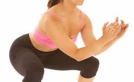
2. Exercise
- Reduce knee pain and inflammation.
- Normalise knee joint range of motion.
- Strengthen lower kinetic chain
- Cardio-vascular exercise
- Improve proprioception, agility and balance.
- Improve function
- Use of walking aids as needed
Land-based exercises are ideal for most people and are strongly recommended.
Exercise has also been found to be beneficial for other co-morbidities and overall health. Walking, resistance training, cycling, yoga and Tai Chi are examples of such exercises. An individualised exercise program should be set by a physiotherapist initially, taking into account the patient's goals and hobbies to ensure long term exercise compliance.
Other Interventions
There are various forms of therapeutic interventions that may or may not be helpful for patients with various degrees of evidence to support them:
- Hydrotherapy - this may be particularly helpful if pain is very high and analgesia is not tolerated. It can be useful to build up strength and reduce stiffness around the knee joint in a non-weight bearing position.
- Taping - works to offload the joint similar to bracing, this is useful in the short term.
- Manual therapy - effective to improve ROM
- Massage - may be useful to control pain in some subjects, but this has low evidence to show its effectiveness
- Bracing
- Electrotherapy - such as TENS and muscle stimulation may be used to improve quadricep strength and has some evidence to show it can help with pain reduction.
What does Evidence says
All guidelines recommended the use of exercise and education/self-management as key interventions for OA. NICE recommended the use of manual therapy (manipulation and stretching) as an adjunct to exercise, particularly in people with hip OA; manual therapy was not included within the other two publications (OARSI stated this modality was not included due to insufficient evidence). More information HERE
If you are suffering from knee pain, contact us for a proper diagnosis and treatment plan.
References:
1. https://academic.oup.com/bmb/article/122/1/151/3792465
2. https://www.transcendhealth.com.au/how-can-a-physiotherapist-help-your-osteoarthritis/
3. https://www.physio-pedia.com/Knee_Osteoarthritis

